
Explainer · July 30, 2026 · 5 min · By Beatriz Holmgren
Silent Rupture Surveillance: How Ultrasound and MRI Actually Compare for Silicone Implants
Silicone implant ruptures often produce no symptoms at all. Here is what the imaging evidence says about screening schedules, what each scan can and cannot see, and why the guidance changed in 2020.
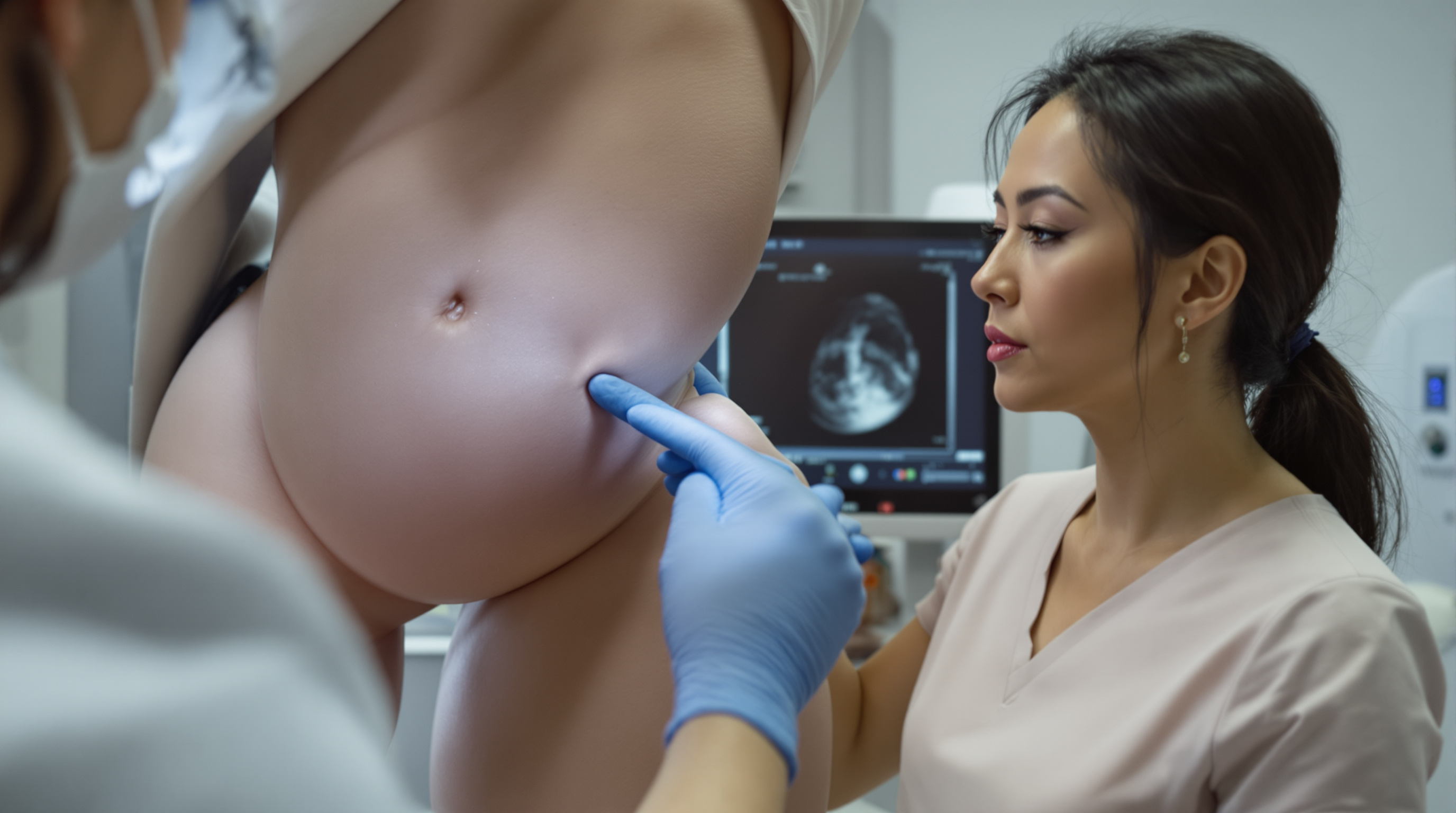
One of the least discussed realities of silicone gel breast augmentation is that a ruptured implant can feel completely normal. The gel in modern devices is cohesive, meaning it holds its shape rather than flowing freely, and the fibrous capsule the body builds around every implant tends to contain the material even after the shell fails. The result is the so called silent rupture: no pain, no visible change, no warning. Population studies using screening MRI have found rupture rates that climb steadily with implant age, which is exactly why regulators recommend imaging surveillance for patients who chose silicone devices, even when everything feels fine.
Saline implants are a different story, and it is worth stating why. When a saline shell fails, the body absorbs the sterile fluid within days and the breast visibly deflates. Detection is essentially self evident, so routine imaging surveillance is not recommended for saline devices. The entire screening conversation below applies to silicone gel implants.
What the current guidance actually says. In 2020, United States regulators updated their labeling recommendations. The prior advice had been MRI at three years after implantation and every two years thereafter, a schedule with famously poor real world compliance because MRI is expensive, time consuming, and often not covered by insurance for asymptomatic screening. The revised guidance recommends either ultrasound or MRI at five to six years post implantation, then every two to three years afterward. If a patient develops symptoms at any point, or if an ultrasound result is equivocal, MRI is the recommended next step.
Why MRI remains the reference standard. MRI detects rupture by visualizing the implant shell and the distribution of silicone directly. Radiologists look for specific signs: the linguine sign, which is the collapsed shell floating inside the gel and appearing as wavy dark lines, indicates intracapsular rupture, where gel remains inside the fibrous capsule. Silicone signal outside the capsule indicates extracapsular rupture. Published sensitivity for MRI in detecting rupture generally falls in the range of roughly 80 to 95 percent, with specificity in a similar band. MRI does not use ionizing radiation, and silicone specific sequences can distinguish gel from fat and water, which is a genuine mechanistic advantage no other modality matches.
What ultrasound can and cannot do. High resolution ultrasound looks for its own signature findings. The stepladder sign, parallel echogenic lines within the implant, is the sonographic cousin of the linguine sign and suggests intracapsular rupture. The snowstorm appearance, a hazy echogenic pattern, suggests free silicone in surrounding tissue. Ultrasound is fast, inexpensive, radiation free, and widely available. Its weaknesses are equally real: accuracy is strongly operator dependent, sensitivity in published series is more variable than MRI, and deep or posterior portions of the implant can be difficult to assess, particularly in patients with larger breasts or significant capsular thickening. In practice, ultrasound functions well as a first pass screen, with MRI reserved for uncertain or positive findings.
What about mammography? Standard mammography is not a rupture screening tool. It can sometimes reveal free silicone that has migrated outside the capsule, but it cannot reliably assess the shell, and intracapsular ruptures are largely invisible to it. Patients with implants should still undergo age appropriate mammographic cancer screening using implant displacement views, sometimes called Eklund views, but that is a separate purpose. Rupture surveillance and cancer screening are two different tasks that happen to involve the same anatomy.
Does a silent rupture demand urgent surgery? This is where nuance matters. An intracapsular rupture with cohesive gel that is fully contained is generally not treated as an emergency, though most surgical societies advise removal, with or without replacement, once rupture is confirmed, because gel can migrate over time and because the diagnosis becomes harder to manage the longer it sits. Extracapsular rupture, where silicone has moved into breast tissue or lymph nodes, is a stronger indication for timely surgery, since migrated silicone can form granulomas, firm inflammatory nodules that complicate both examination and future imaging.
Practical takeaways. First, ask what generation of device you have and when it was placed, because rupture risk is a function of implant age and shell design. Second, put the surveillance schedule on an actual calendar: first scan at five to six years, then every two to three years. Third, understand the trade: ultrasound is accessible and reasonable as a screening tool, MRI is more definitive and is the appropriate follow up when anything looks ambiguous. Fourth, do not interpret a normal feeling breast as evidence of an intact implant. The entire rationale for surveillance is that the shell can fail quietly.
None of this should be read as alarming. Modern cohesive gel devices are more durable than earlier generations, and rupture is a manageable, well understood event. The point of screening is simply to convert an invisible mechanical failure into a known quantity, so decisions about removal or replacement can be made on a schedule the patient controls rather than one dictated by a late complication.
Related reading: Saline vs. silicone implants: the core difference.