
Field Notes · July 26, 2026 · 6 min · By Rohan Chatterton
Implant Rippling: the Pinch Test That Predicts It and What Actually Reduces It
Rippling is not a manufacturing defect and not usually a surgical error. It is a coverage problem, and the amount of coverage you have is a number your surgeon measures with two fingers in the first five minutes of a consult.
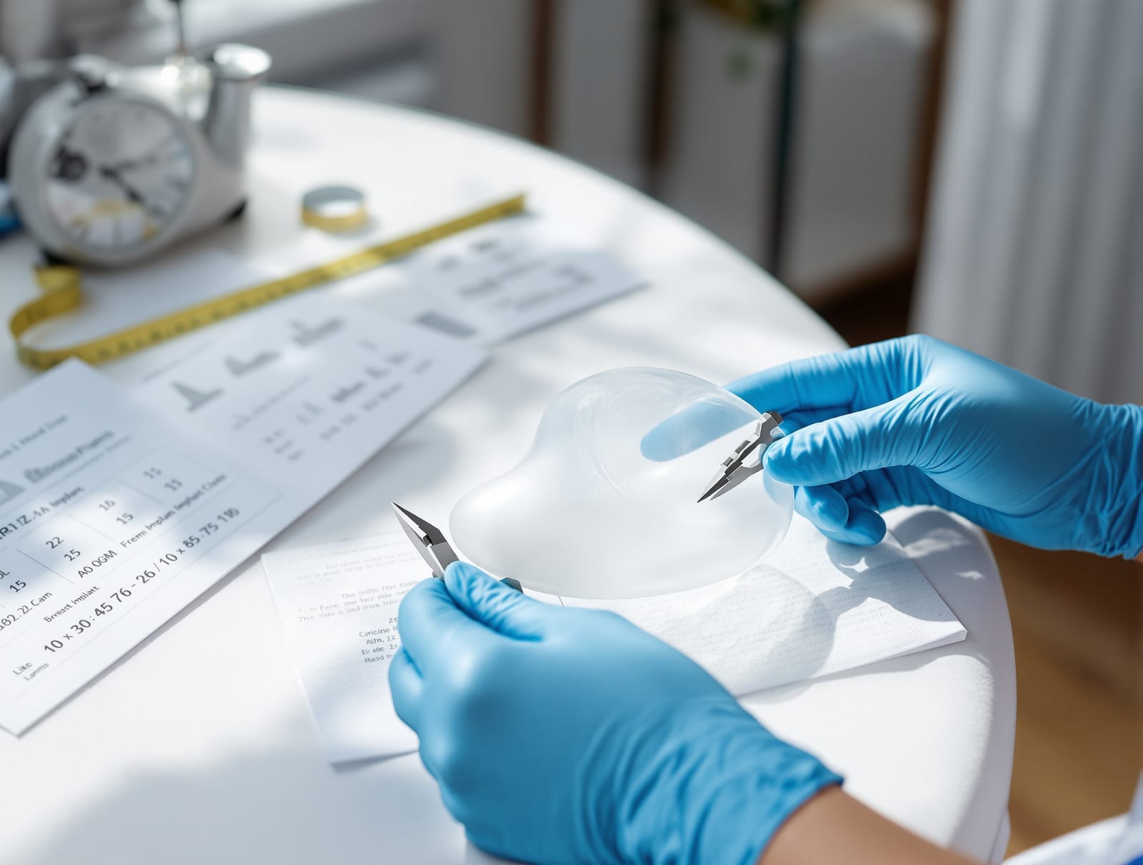
Rippling is the complaint that arrives late. Not in the first month, when everything is swollen and firm, but somewhere in the second year, usually noticed while leaning forward in a mirror or after a stretch of weight loss. A faint corrugation along the outside of the breast, or a set of fine vertical lines at the cleavage, visible in certain light and certain positions and invisible in others.
Patients tend to interpret it as something having gone wrong. It is more accurate to say that something was always going to be visible, and the only real question was how much tissue was standing between the implant surface and the light.
The original element in this piece is a four site pinch thickness self check with an interpretation key, drawn from the soft tissue measurements that implant selection systems have used for over two decades but that patients are almost never taught to take themselves. It takes two minutes. It will not replace your surgeon's measurement, and the reasons it will not are stated plainly at the end.
What rippling physically is. An implant is a filled shell. Where the shell is not fully supported, it folds, in the same way a partly filled water bottle creases. Saline devices ripple more readily than silicone because water has no cohesion of its own and the shell carries the shape. Cohesive silicone gel resists folding in proportion to how firmly it is cross linked. But no filler prevents a fold from showing if the tissue over it is thin enough, and that is the part patients underestimate. Rippling is a visibility problem before it is a device problem.
Which is why the measurement that predicts it is a measurement of you, not of the implant. Soft tissue pinch thickness at the upper pole has been part of formal implant selection since the tissue based planning systems published in Plastic and Reconstructive Surgery in the early 2000s, which built device choice around measured patient tissue characteristics rather than around a requested cup size (Plastic and Reconstructive Surgery, 2002). The logic has not changed. Thin cover means the device must be placed deeper, and deeper means under muscle.
The four site pinch check. Do this standing, in front of a mirror, in a warm room, on an unoperated chest or on an augmented one. You are pinching a fold of skin and fat between thumb and index finger, lifting gently, and estimating the thickness of the doubled fold. Then halve that number, because a pinch measures two layers.
Site one, the upper pole. Two finger widths below the collarbone, in the vertical line of the nipple. This is the site the planning systems care about most, because it is where the top edge of an implant sits and where edge visibility and upper pole rippling appear.
Site two, the medial border. Just lateral to the sternum at the level of the nipple, the cleavage zone. This is the site that determines whether a fold shows in a low neckline, which is the presentation people find most bothersome.
Site three, the lateral border. Along the side of the breast toward the armpit, where a well fitted bra band crosses. Lateral rippling is the version most often noticed in profile and when leaning.
Site four, the lower pole. Just above the fold beneath the breast. This tissue is usually thickest and rarely the problem, so it functions as your internal control. If your lower pole pinch is not clearly thicker than your upper pole pinch, you are a thin cover patient across the board.
The interpretation key. Halve each doubled fold to get single layer thickness. An upper pole single layer thickness of roughly one centimeter or more, which is a doubled pinch of about two centimeters or more, is the range in which planning systems have historically considered subglandular placement viable without predictable edge visibility. Below that, the same systems push toward submuscular or dual plane coverage, precisely because the muscle is being recruited as additional cover rather than for any structural reason. If your medial pinch is markedly thinner than your upper pole pinch, you are the patient for whom a wider, higher projection device placed close to the midline is most likely to produce visible cleavage rippling, a pattern addressed directly in published guidance on preventing edge visibility with high and extra high profile round implants (Aesthetic Plastic Surgery, 2019).
That last point is the counterintuitive one and it is worth stating as bluntly as possible. Going bigger does not hide rippling. It stretches the same tissue over a larger surface, thins it further, and increases the fold length available to be seen. The relationship between projection, base width and cover is the practical content of implant profiles and projection explained and of how implant size actually gets chosen.
What actually reduces it. Three things, in descending order of evidence. Placing the device under the muscle, which adds a layer of cover at the upper pole where it matters most. Choosing a more cohesive gel rather than saline, which resists folding at the shell level, discussed further in saline vs silicone implants and cohesive gel and implant shapes. And in revision cases, adding soft tissue support, where absorbable mesh scaffolds have been reported as an adjunct in revision augmentation specifically to address soft tissue thinning and edge visibility (Aesthetic Surgery Journal, 2023).
Two things that are frequently offered and do far less. Fat grafting over the implant genuinely adds cover, but graft retention is variable and it adds an operation. Massage does nothing for rippling at all, because rippling is not a capsule phenomenon.
What the studies do not tell you. There is no validated patient administered version of the pinch test, and no study has correlated self measured pinch thickness with rippling incidence. The thresholds above come from surgical planning systems applied by trained hands with the patient positioned deliberately, and a self pinch will typically over read, because it is easy to catch more tissue than intended. Nor is there a good prospective study of rippling incidence in cosmetic augmentation stratified by cover thickness, which is a remarkable gap given how long the mechanism has been understood. What exists is mechanistic reasoning plus surgeon series, and it is enough to plan with but not enough to promise with.
So use the check for what it is good for. It is not a prediction. It is a way of arriving at a consult already knowing whether you are a thin cover patient, so that when a surgeon recommends the muscle plane you understand that the recommendation is about your tissue and not about their preference. The FDA maintains a general patient resource on implant risks and long term monitoring at FDA breast implants, which is worth reading alongside any device conversation.
One quotable line to take into the room: rippling is not caused by the implant you chose, it is revealed by the tissue you have, and the only variable a bigger implant reliably changes is how much of it there is to see.