
Explainer · July 27, 2026 · 5 min · By Beatriz Holmgren
Over, Under, or Dual Plane: How Implant Pocket Placement Actually Changes Outcomes
The plane your surgeon chooses for the implant pocket affects animation, contracture risk, mammography, and how the result ages. Here is what the anatomy actually dictates.
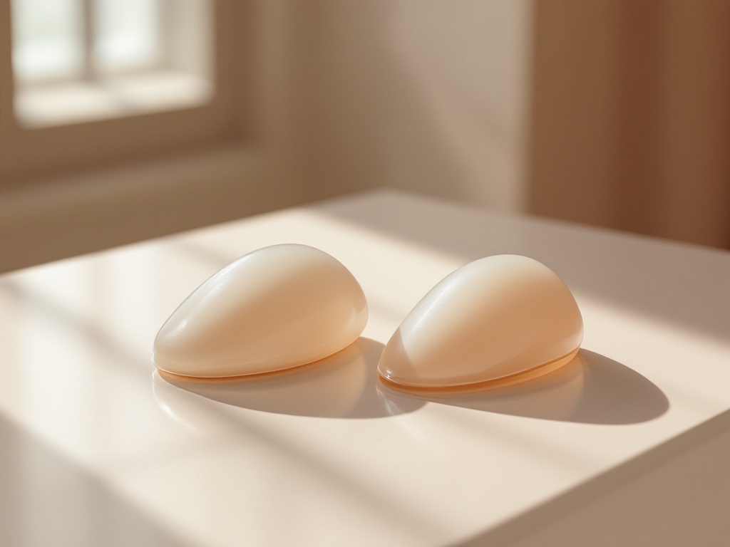
When patients research breast augmentation, most of the attention goes to implant type and size. But surgeons often say the more consequential decision is the pocket: whether the implant sits above the pectoralis major muscle, below it, or in a hybrid position called the dual plane. Each option changes the mechanics of how the implant is covered, how it moves, and how the breast behaves over years, not weeks.
Subglandular placement puts the implant directly behind the breast gland and in front of the pectoralis major. The appeal is straightforward. The muscle is untouched, so early recovery tends to be less painful, and the implant is not compressed or displaced when the pec contracts. In patients with a mild degree of glandular droop, a subglandular implant can fill the loose envelope more directly, sometimes avoiding a lift. The trade-off is coverage. The only tissue between the implant and the skin is gland and fat. In slim patients with thin soft tissue, this raises the odds of visible implant edges, palpable rippling along the upper and inner breast, and a rounder, more obviously augmented upper pole. Pinch thickness at the top of the breast, often cited around 2 centimeters as a rough threshold, is one way surgeons screen for whether subglandular coverage is adequate.
Submuscular placement, more precisely called subpectoral or partial submuscular, slides the upper portion of the implant beneath the pectoralis major. Total muscle coverage is rare in cosmetic augmentation; the lower pole of the implant usually sits below the muscle's edge, covered by gland and fascia. The muscle adds a living layer of padding over the upper implant, which softens the transition from chest wall to breast and reduces visible rippling where it matters most cosmetically. There is also a mechanistic argument on capsular contracture: multiple large series have reported lower contracture rates with submuscular pockets, plausibly because muscle motion massages the capsule and because the pocket has less direct contact with ductal tissue, a potential source of low-grade bacterial contamination that is one leading theory behind contracture.
The cost of going under the muscle is animation deformity. Because the pectoralis is cut along its lower attachments to make room, contracting the muscle, as in pushing, lifting, or certain gym movements, can visibly distort or flatten the implant. For most patients this is a minor cosmetic quirk. For athletes, bodybuilders, and people whose work involves repetitive pressing motions, it can be a genuine functional and aesthetic complaint, and it is one of the main reasons some of these patients choose subglandular or subfascial pockets despite thinner coverage.
Dual plane technique, described in the surgical literature in the early 2000s, is the compromise that has become the default in many practices. The upper implant sits under the muscle for coverage; the lower implant sits under the gland, because the surgeon releases the muscle's lower border and, depending on the variant, some of the attachments between muscle and gland. This lets the implant expand the lower pole and pick up mild sagging while keeping muscle padding over the visible upper edge. Dual plane is graded, commonly one to three, based on how much release is performed, with higher grades used for patients with more glandular ptosis or constricted lower poles.
A few practical implications follow from the mechanics. On mammography, submuscular and dual plane placement generally allow better visualization of breast tissue, because the implant can be displaced away from the gland during imaging. Radiologists use special displacement views either way, but thin patients with subglandular implants tend to have more tissue obscured. On long-term aging, subglandular implants tend to descend with the gland, which some patients prefer because breast and implant stay together. Submuscular implants can stay higher while natural tissue drapes off the front over time, a pattern sometimes called a waterfall or Snoopy deformity, which is one reason dual plane release matters in patients with any laxity at the start.
What does the evidence not support? The idea that one pocket is universally superior. Contracture data favor submuscular positioning on average, but modern practices such as careful pocket irrigation, minimal implant handling, and inframammary incisions have narrowed the gap. Recovery differences are real but temporary, usually measured in days to a couple of weeks of extra soreness with muscle release. And no pocket choice compensates for an implant that is too wide or too heavy for the patient's chest, which drives thinning of tissue and bottoming out regardless of plane.
The useful takeaway for a consultation is to ask three questions. How thick is my upper pole tissue, in the surgeon's own measurement? How active is my pectoralis in daily life and training? And how much natural droop needs to be addressed now versus managed later? The answers, more than any brochure, point to the right plane.
Related reading: Over, Under, or In Between: How Implant Plane Actually Changes Outcomes.