
Explainer · July 27, 2026 · 5 min · By Beatriz Holmgren
Over, Under, or In Between: How Implant Pocket Placement Actually Changes Outcomes
Subglandular, submuscular, and dual plane placement are often presented as a matter of surgeon preference. In reality, each pocket changes soft tissue coverage, animation, mammography, and long-term revision risk in measurable ways.
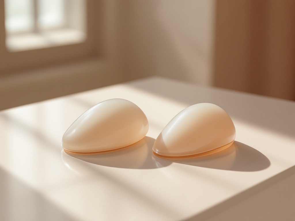
Ask ten patients where their breast implant sits and most will say behind the breast. The more useful question is behind which layer. The pocket a surgeon creates, meaning the anatomical space where the implant lives, is one of the strongest predictors of how an augmentation looks at year one, how it ages at year ten, and what kinds of complications are more or less likely. There are three main options: subglandular (over the pectoralis major muscle, under the breast gland), submuscular (under the muscle, more accurately called subpectoral in most modern practice), and dual plane, a hybrid in which the upper portion of the implant sits under the muscle while the lower portion sits under the gland.
Why coverage is the core variable. An implant is only as concealed as the tissue on top of it. Surgeons commonly assess this with a pinch test at the upper pole of the breast. When the pinched tissue measures less than roughly 2 centimeters, a subglandular implant tends to show its edges over time. That shows up as visible rippling, a hard shelf at the upper border, and a rounded, obviously augmented contour. Placing the implant under the pectoralis major borrows muscle thickness to pad the upper pole, which is why thinner patients are usually steered toward subpectoral or dual plane placement. Patients with more of their own breast tissue have more flexibility, and subglandular placement can produce a natural result with a faster recovery.
The trade-off nobody escapes: animation. Muscle coverage comes at a price. When the pectoralis contracts, during a push-up, a chest press, or even reaching across the body, a subpectoral implant can visibly shift, flatten, or distort. This is called animation deformity. For most patients it is a minor cosmetic quirk. For athletes, bodybuilders, and anyone whose sport loads the chest heavily, it can be a genuine functional and aesthetic problem. Subglandular placement eliminates animation entirely because the muscle never touches the implant. This is one reason some very active patients with adequate tissue coverage deliberately choose the over-the-muscle pocket despite its other drawbacks.
Capsular contracture risk differs by pocket. Every implant develops a scar capsule. In a minority of patients that capsule tightens and hardens, a complication called capsular contracture. Pooled data across decades of studies consistently show lower contracture rates with submuscular placement than subglandular. The proposed mechanisms are plausible: the muscle may act as a barrier reducing bacterial contamination from the ducts of the breast gland, and constant muscle movement may mechanically discourage a thick, contracted capsule from forming. Contracture still happens in every pocket, but the risk gradient is real and worth factoring in, particularly for patients planning to keep implants for decades.
Mammography and screening. Implants block a portion of breast tissue from view on standard mammograms regardless of position, which is why augmented patients receive additional displacement views. That said, submuscular implants generally allow more complete visualization of the gland because the implant sits behind it rather than pressed into it. For patients with elevated breast cancer risk or dense tissue, this is a legitimate conversation to have before surgery, not after.
Where dual plane fits. Dual plane placement was developed to solve a specific problem: patients who need upper pole muscle coverage but whose lower breast tissue is slightly loose or droopy. By releasing the lower edge of the pectoralis, the surgeon lets the implant expand the lower pole directly against the gland while keeping muscle over the top edge. Mechanically, this allows the implant to fill mild sagging without a lift in carefully selected cases, while still hiding the implant border where tissue is thinnest. It has become the default approach in many practices precisely because it hedges between the two extremes, though it does not eliminate animation and it requires more technical precision than either pure pocket.
Recovery differences are real but temporary. Subglandular surgery avoids cutting or lifting muscle, so early pain is typically milder and return to normal activity is faster. Subpectoral and dual plane patients often describe more pressure and tightness in the first one to two weeks as the muscle stretches. By six weeks, recovery experiences largely converge, so recovery alone is a weak reason to choose a pocket that fits the anatomy poorly.
The honest summary. There is no universally superior pocket. Thin tissue pushes the decision toward muscle coverage. Heavy chest athletics push it away. Contracture history, screening needs, degree of sag, and implant size all move the needle. A surgeon who recommends the same pocket for every patient is using a template, not an assessment. The most productive question to bring to a consultation is not which pocket is best, but which pocket best matches this tissue, this lifestyle, and this timeline, and why.
Related reading: Over, Under, or In Between: How Implant Plane Actually Changes Outcomes.