
Explainer · July 28, 2026 · 5 min · By Beatriz Holmgren
Over, Under, or In Between: How Implant Pocket Placement Actually Works
Subglandular, submuscular, and dual plane placement each change how an implant looks, feels, and ages. Here is the mechanical logic behind each pocket, minus the marketing.
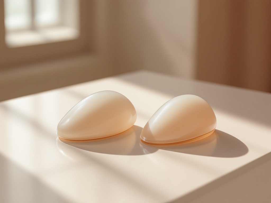
Ask three surgeons where a breast implant should sit and you may get three different answers. That is not because anyone is wrong. It is because pocket placement is a trade-off problem, and the right answer depends on tissue thickness, lifestyle, and what a patient is willing to accept long term. This explainer walks through the three main options, the mechanisms behind each, and the questions worth asking in a consultation.
The anatomy in one paragraph. The breast gland sits on top of the pectoralis major, the fan-shaped chest muscle. Between the gland and the muscle is a natural plane of loose tissue. An implant can be placed above the muscle in that plane (subglandular), fully or partially beneath the muscle (submuscular), or in a hybrid position where the upper portion is covered by muscle and the lower portion sits directly under the gland (dual plane). Some surgeons also describe a subfascial pocket, where the implant sits under the thin fibrous sheet covering the muscle but not under the muscle itself.
Subglandular: closest to the natural breast position. Placing the implant on top of the muscle puts it where breast tissue naturally lives, which can produce a fuller upper pole and more direct projection. Recovery tends to be faster because the muscle is not lifted or divided, and there is no animation deformity, the visible distortion that can occur when a submuscular implant moves during pectoral contraction. The trade-offs are mechanical. With only skin and gland covering the device, thin patients are more likely to see rippling, feel implant edges, or show visible transitions at the top of the implant. Several long-running datasets also report higher rates of capsular contracture in subglandular pockets, possibly because the implant sits closer to milk ducts and their bacterial flora, one of the leading theories behind contracture formation.
Submuscular: more coverage, more compromise with the muscle. Sliding the implant under the pectoralis major adds a layer of living tissue over the upper implant. That extra padding softens edges, reduces visible rippling, and tends to lower contracture rates, likely because constant muscle motion gently massages the pocket and because the implant is separated from the ductal system. Mammographic imaging is also somewhat easier to interpret with the implant behind the muscle. The costs: recovery involves more early discomfort because muscle fibers are stretched or partially released, and animation deformity is possible. For athletes who train the chest heavily, repeated pectoral contraction can gradually displace implants outward or downward over years, a phenomenon sometimes called lateral drift.
Dual plane: the compromise most commonly used today. In a dual plane approach, the surgeon releases the lower attachment of the pectoralis so the upper implant is muscle-covered while the lower implant sits directly behind the gland. The mechanism is straightforward: coverage where tissue is thinnest (the upper pole, where rippling shows most) and direct implant-to-gland contact where the breast needs to expand and redrape (the lower pole). This is particularly useful for patients with mild sagging or a tight lower breast fold, because the implant can help shape the lower pole rather than sitting behind a muscle that resists expansion. Surgeons grade dual plane from type 1 to type 3 depending on how much muscle is released, tailoring coverage to the individual chest.
What actually drives the decision. The most important variable is the pinch test: how much soft tissue exists in the upper breast. Under roughly 2 centimeters of pinch thickness, most surgeons favor muscle coverage or a highly cohesive implant, because thin tissue cannot camouflage the device. Other factors include planned pregnancies, weightlifting habits, existing sag, implant size (larger devices show edges more readily), and whether the patient prioritizes a fast recovery or long-term coverage.
A note on subfascial placement. The subfascial pocket has gained interest as a middle path: no muscle division, so no animation deformity, but slightly more support than a pure subglandular pocket. The fascia itself is thin, typically under a millimeter, so its camouflage effect is modest. Evidence remains less extensive than for the other pockets, and results depend heavily on the patient having adequate native tissue.
Questions worth bringing to a consultation. Ask which pocket the surgeon recommends and why, specifically what your tissue measurements show. Ask how they handle animation deformity if you lift weights. Ask what a revision would involve, since switching pockets later is possible but adds complexity. And ask about contracture rates in their own practice, not just published averages.
The bottom line. No pocket is universally superior. Subglandular offers easier recovery and natural motion at the cost of coverage. Submuscular offers padding and lower contracture risk at the cost of animation and a harder early recovery. Dual plane blends both, which is why it has become the default for many surgeons. The best predictor of a good outcome is not the pocket itself but whether the pocket matches the tissue it has to work with.
Related reading: Over, Under, or In Between: How Implant Plane Actually Changes Outcomes.