
Explainer · July 28, 2026 · 6 min · By Beatriz Holmgren
Over, Under, or In Between: How Implant Pocket Placement Actually Works
Subglandular, submuscular, and dual plane placement change how an implant looks, feels, and ages. Here is the anatomy behind each option, and the tradeoffs surgeons weigh when choosing one.
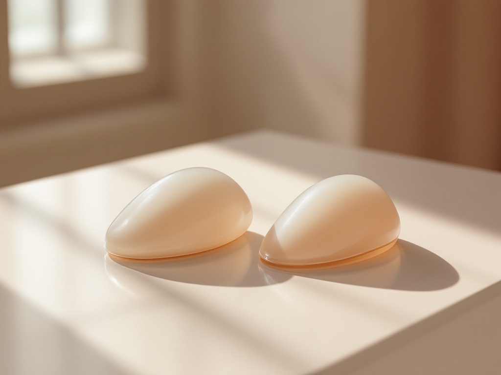
When patients research breast augmentation, they usually focus on implant size and fill material. Surgeons, however, spend just as much time deciding where the implant will sit relative to the pectoralis major, the fan-shaped chest muscle that covers much of the upper breast. That decision, known as pocket placement, influences upper pole contour, rippling risk, animation deformity, recovery time, and even long-term complication rates. Understanding the three main options makes consultations far more productive.
Subglandular placement: on top of the muscle. In a subglandular pocket, the implant sits between the breast gland and the pectoralis major. The mechanical logic is simple: the implant pushes the natural breast tissue forward without any muscular interference. Recovery tends to be faster because the muscle is not lifted or released, and there is no risk of animation deformity, the visible distortion that can occur when a muscle contracts over an implant. The tradeoff is coverage. Only skin and gland separate the implant from the surface, so in patients with thin soft tissue, the upper edge of the implant can be visible, and rippling is more likely with saline or lightly filled silicone devices. Some studies have also associated subglandular placement with higher rates of capsular contracture, possibly because the pocket sits closer to ductal tissue and its bacterial flora, a factor in the biofilm theory of contracture.
Submuscular placement: under the muscle. Here the surgeon elevates the pectoralis major and positions the implant beneath it. In practice, total muscle coverage is rare, since the pectoralis does not extend over the entire lower breast. What most people call submuscular is really partial coverage of the upper implant. The mechanism behind its popularity is padding: an extra layer of muscle softens the transition at the upper pole, reduces visible rippling, and appears to lower capsular contracture rates in several large series. Mammographic imaging is also somewhat easier to interpret with muscle between the gland and the implant. The costs are real, though. Recovery involves more discomfort in the first week because a muscle has been stretched. And because the pectoralis remains attached and functional, flexing it can compress or shift the implant, producing animation deformity that ranges from subtle to obvious depending on the patient and the dissection.
Dual plane: the hybrid most commonly used today. Dual plane placement splits the difference. The surgeon releases the lower attachments of the pectoralis major so that the upper portion of the implant sits under muscle while the lower portion sits directly behind the gland. Mechanically, this does two useful things at once. The muscle still pads the upper pole, where thin tissue and rippling are most visible, while the lower pole is free to expand and redrape over the implant. That lower pole freedom matters for patients with mild sagging or a constricted lower breast, because the implant can fill and shape tissue that a fully submuscular pocket would restrain. Surgeons describe dual plane in grades, typically one to three, based on how far the muscle is released. Higher grades allow more glandular redraping for patients with looser or lower-sitting tissue.
How surgeons actually choose. The single most important variable is soft tissue thickness, often assessed with a pinch test at the upper breast. A common threshold cited in surgical literature is roughly two centimeters: patients with less coverage than that generally benefit from muscle over the upper implant, while patients with generous natural tissue can consider subglandular placement with fewer visibility concerns. Lifestyle matters too. Competitive athletes and bodybuilders who use the pectoralis heavily may prefer subglandular placement to avoid animation deformity, accepting the coverage tradeoff. Implant type interacts with the decision as well, since highly cohesive silicone devices ripple less than saline and can tolerate thinner coverage.
What the evidence supports, and what it does not. Reported data consistently link partial muscle coverage with lower capsular contracture rates and better upper pole camouflage in thin patients. Data also consistently show that animation deformity is essentially exclusive to pockets involving muscle, and that subglandular recovery is shorter. What the evidence does not support is the idea that one pocket is universally superior. Revision surgery sometimes involves converting from one plane to another precisely because the original choice did not match the patient's tissue or lifestyle.
The practical takeaway. Pocket placement is not a preference item like choosing a size, it is an anatomical decision driven by measurable factors: tissue thickness, degree of sagging, muscle use, and implant characteristics. A useful consultation question is not simply which plane a surgeon prefers, but why a particular plane suits your specific pinch thickness and breast shape. Patients who understand the mechanics of over, under, and in between are better equipped to weigh recovery, appearance, and long-term risk on their own terms.
Related reading: Over, Under, or In Between: How Implant Pocket Placement Actually Changes Outcomes.