
Explainer · August 2, 2026 · 5 min · By Beatriz Holmgren
Over, Under, or In Between: How Implant Pocket Placement Actually Changes Outcomes
Subglandular, submuscular, and dual plane placement are not interchangeable options. Here is what each pocket does to the tissue, the implant, and the long-term result.
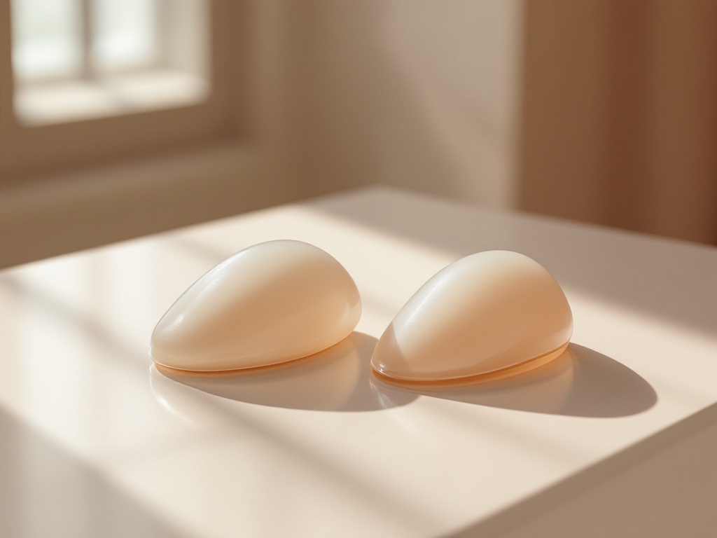
Ask three surgeons where a breast implant should sit and you may get three different answers. That is not because the field is confused. It is because pocket placement is a trade-off decision, and the right answer depends on tissue thickness, activity level, and how a patient's breast is expected to age. This explainer breaks down the three main pockets, the mechanisms behind each, and where the evidence actually points.
The anatomy in plain terms
The breast gland sits on top of the pectoralis major, the fan-shaped chest muscle. An implant can be placed above that muscle and directly behind the gland (subglandular), fully or mostly beneath the muscle (submuscular), or in a hybrid position where the upper portion of the implant is covered by muscle and the lower portion sits under gland only (dual plane). A fourth variant, subfascial placement, positions the implant under the thin connective tissue layer covering the muscle but not under the muscle itself.
Subglandular: direct, but demanding of tissue
Placing the implant behind the gland is the most anatomically direct approach. The implant pushes the breast forward from immediately behind it, which can produce a fuller, rounder upper pole and allows the implant to move naturally with the breast. Recovery tends to be faster because no muscle is released or stretched.
The trade-off is coverage. Everything separating the implant from the outside world is skin, fat, and gland. In patients with thin tissue, typically measured as a pinch test under about two centimeters at the upper pole, this can mean visible implant edges, rippling, and a step-off where the implant begins. Mechanistically, rippling occurs because the implant shell folds slightly under gravity, and thin overlying tissue cannot mask those folds. Multiple studies also associate subglandular placement with higher rates of capsular contracture, the scar tissue tightening that can distort or harden the breast. One proposed mechanism is proximity to the ductal system of the gland, which harbors low levels of bacteria that may contribute to biofilm formation on the implant surface.
Submuscular: more coverage, more mechanics
Placing the implant under the pectoralis major adds a layer of living, vascularized muscle over the upper implant. This does three useful things. It softens the transition at the upper pole so the implant edge is less visible. It appears to lower capsular contracture rates, possibly by keeping the implant away from glandular bacteria and by the massaging effect of muscle motion. And it improves mammographic imaging, since the implant is pushed further from breast tissue during compression views.
The costs are mechanical. Recovery involves more discomfort because the muscle is partially released from its lower attachments and then stretched over the implant. More importantly, the muscle keeps working after surgery. When the pectoralis contracts, it can compress or displace the implant, a phenomenon called animation deformity. For most patients this is a subtle flicker during exercise. For competitive athletes or heavy lifters, it can be noticeable and bothersome, and over years, repeated muscle pressure can push implants outward or downward.
Dual plane: the current default for a reason
Dual plane placement, described in the surgical literature in the early 2000s, attempts to split the difference. The upper half to two thirds of the implant sits beneath the muscle, gaining coverage and the contracture benefit where tissue is thinnest. The lower portion sits behind the gland only, allowing the implant to expand the lower pole and letting the gland redrape over it. Surgeons adjust how much muscle is released, often described as dual plane types one through three, based on how much the natural breast sags and how tightly the gland adheres to the muscle.
This is why dual plane has become the most common approach in many practices. It handles the widest range of anatomies, including mild sagging that a fully submuscular pocket would not correct well, because a fully covered implant can end up sitting high while the natural gland slides down in front of it, a mismatch sometimes called a waterfall deformity.
What actually drives the decision
Four factors matter most. First, tissue pinch thickness at the upper pole: thin tissue pushes the decision toward muscle coverage. Second, activity profile: heavy chest-focused training argues for subglandular or subfascial placement to avoid animation issues. Third, degree of existing ptosis, or sag: more sag favors dual plane or a combined lift. Fourth, implant characteristics: cohesive silicone gel ripples less than saline, which gives thin-tissued patients slightly more flexibility.
The honest bottom line
No pocket is universally superior. Subglandular placement trades coverage for simplicity and muscle independence. Submuscular placement trades recovery comfort and animation for camouflage and lower contracture risk. Dual plane blends both and fits the most anatomies, which explains its dominance, but it is not automatic. A consultation that skips tissue measurements and jumps straight to a recommended pocket is a consultation worth questioning. The pocket should be chosen to fit the tissue, not the surgeon's routine.
Related reading: Above, Below, or Dual Plane: How Implant Pocket Placement Actually Works.