
Explainer · July 29, 2026 · 4 min · By Beatriz Holmgren
Above, Below, or Dual Plane: How Implant Pocket Placement Actually Works
Subglandular, submuscular, and dual plane placement are often presented as a menu of preferences. In reality, each pocket changes how an implant behaves over years, not just how it looks in week one. Here is the mechanism-level breakdown.
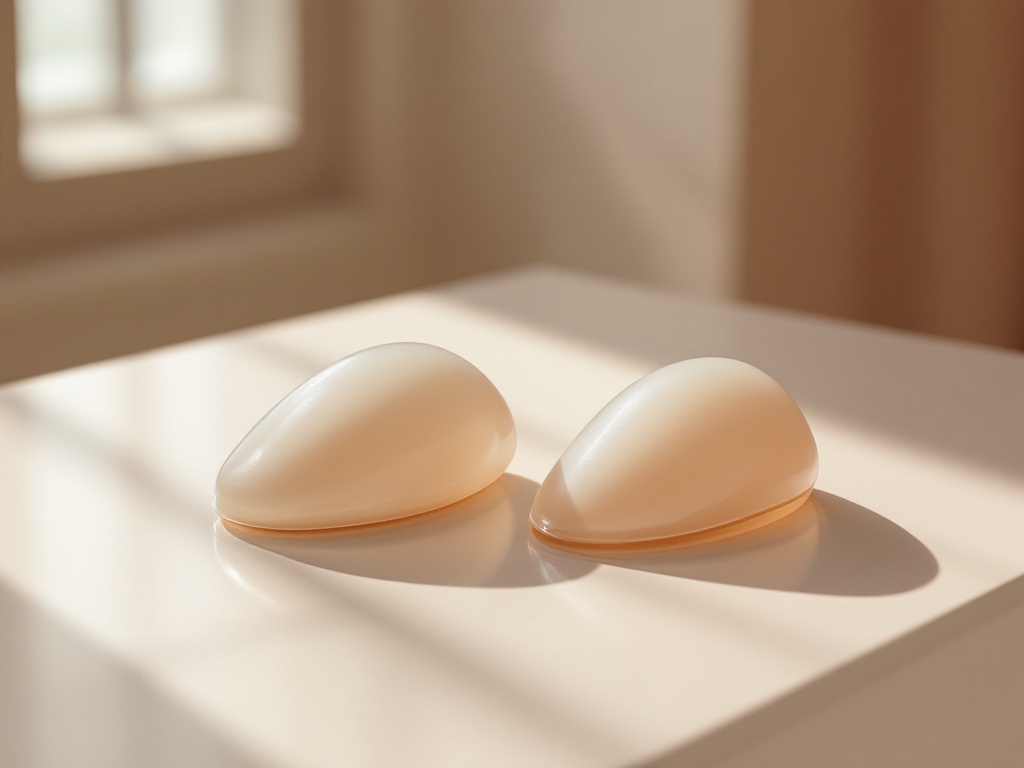
Ask three surgeons where a breast implant should sit and you may get three confident answers. That is not because the science is unsettled. It is because pocket selection is a matching problem: the right plane depends on tissue thickness, breast shape, activity level, and how a patient's chest is likely to age. Understanding the mechanics makes consultations far more productive.
The three pockets, defined. In subglandular placement, the implant sits between the breast gland and the pectoralis major muscle. In submuscular or subpectoral placement, the upper portion of the implant sits beneath that muscle. Dual plane, the most common approach in modern practice, is a hybrid: the upper pole of the implant is covered by muscle while the lower pole sits directly behind the gland, because the surgeon releases the muscle's lower attachments to let the implant settle into the lower breast.
Why coverage matters more than marketing. The central variable is soft tissue thickness. An implant is a smooth or textured silicone shell, and anything that separates it from the skin softens its edges visually and to the touch. A patient with a pinch test of less than roughly two centimeters in the upper pole has thin coverage, and a subglandular implant in that setting is more likely to show visible rippling, palpable edges, and a rounded, obviously augmented upper contour. Adding the pectoralis muscle on top of the implant adds several millimeters of living padding exactly where coverage is thinnest.
The case for subglandular. In patients with generous natural tissue, subglandular placement has real advantages. Recovery tends to be faster and less painful because no muscle is elevated. There is no animation deformity, the visible distortion of the implant when the pectoralis contracts, which matters to weightlifters, climbers, and athletes. The implant also follows the gland more directly, which can suit patients with mild sagging where a submuscular implant might sit high while the natural tissue slides off it, a mismatch surgeons call a waterfall or Snoopy deformity.
The case for submuscular and dual plane. Muscle coverage does more than hide edges. Mammography is generally easier to interpret with submuscular implants because more glandular tissue can be imaged without the implant obscuring it. Multiple large series have also associated submuscular placement with lower rates of capsular contracture, the hardening of scar tissue around the implant. The proposed mechanisms include better vascularized coverage and reduced bacterial contamination from breast tissue, though contracture is multifactorial and no pocket eliminates the risk.
Why dual plane became the default. Full submuscular placement, where muscle and fascia cover the entire implant, constrains the lower pole and can flatten the natural shape of the breast. Dual plane was developed to solve this. By releasing the muscle inferiorly, the surgeon lets the implant expand the lower pole and fill mild droop while keeping muscle over the visible and palpable upper pole. Surgeons adjust how much release to perform, often described as dual plane types one through three, depending on how loose or ptotic the tissue is. In effect, dual plane trades a small amount of animation risk for better shape control in a wide range of anatomies.
Trade-offs patients should actually weigh. Submuscular and dual plane recoveries involve more early discomfort and tightness because muscle has been elevated, and implants typically take longer to settle. Animation deformity is real, though usually mild, and matters most to people who train chest muscles hard. Subglandular placement carries higher long-term risks of visible rippling as tissue thins with age and weight change, and thin patients who choose it sometimes need fat grafting later to camouflage edges. Neither pocket is reliably better for nipple sensation, which depends more on implant size and dissection than on plane.
Myth worth retiring: the pocket determines cup size or projection. It does not. Volume, base width, and implant profile drive size and projection. The pocket determines how naturally that volume integrates with your tissue and how it holds up over a decade.
Questions to bring to a consultation. Ask what your upper pole pinch measurement is and how it informed the recommendation. Ask whether a dual plane approach was considered and at what level of release. Ask how the plan accounts for your sport or profession if pectoral animation would bother you. And ask what a revision would look like in ten years, because pocket choice today shapes the options later: converting planes is possible but adds complexity.
The honest summary is that dual plane earned its dominance by balancing coverage, shape, and contracture data for the average anatomy. Subglandular remains a legitimate choice for well-padded, athletic patients who prioritize natural movement and fast recovery. The best decision is not the trendiest pocket. It is the one matched to the tissue you actually have.
Related reading: Over, Under, or In Between: How Implant Pocket Placement Actually Works.