
Explainer · July 31, 2026 · 5 min · By Beatriz Holmgren
Over, Under, or In Between: How Implant Pocket Placement Actually Works
Subglandular, submuscular, and dual plane are more than menu options. Each pocket changes how an implant looks, feels, and ages. Here is the anatomy behind the choice.
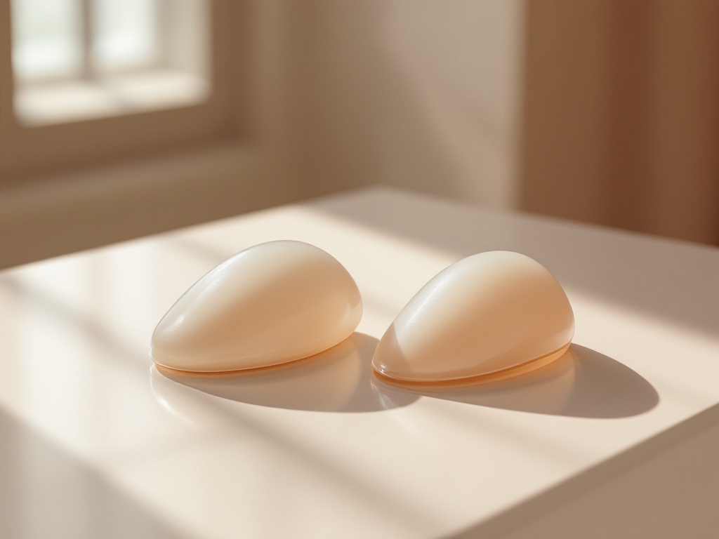
Ask three surgeons where a breast implant should sit and you may get three different answers. That is not because the field is confused. It is because pocket placement is a genuine trade-off, and the right answer depends on tissue thickness, lifestyle, and what a patient is willing to accept long term. This explainer walks through the three main pockets, what each one does mechanically, and why the dual plane approach has become the default for many primary augmentations.
The anatomy in one paragraph. The breast gland sits on top of the pectoralis major, a fan-shaped chest muscle. Between the gland and the muscle is a thin layer of fascia. An implant can be placed above the muscle, directly behind the gland (subglandular), fully or mostly behind the muscle (submuscular), or in a hybrid position where the upper portion of the implant sits under muscle while the lower portion sits under gland (dual plane). A less common variant, subfascial placement, tucks the implant under the fascia but above the muscle fibers.
Subglandular: direct projection, less coverage. Placing the implant behind the gland is mechanically simple. The implant pushes the breast tissue forward, recovery tends to be faster because muscle fibers are not divided or stretched, and there is no risk of animation deformity, the visible distortion that can happen when a flexed pectoral muscle presses on an implant. The cost is coverage. In the upper pole of the breast, the only soft tissue disguising the implant edge is skin and gland. Patients with a pinch thickness under roughly two centimeters in the upper pole are more likely to show rippling, visible implant edges, and a rounded, obviously augmented upper contour. Multiple studies also associate subglandular placement with somewhat higher rates of capsular contracture, the scar tissue tightening that can harden and distort the breast, possibly because the pocket sits closer to ductal bacteria.
Submuscular: coverage at a cost. Putting the implant behind the pectoralis major borrows muscle thickness to camouflage the upper edge of the implant. This produces a softer, more gradual slope in the upper pole and makes rippling less visible in thin patients. It also tends to give clearer mammogram imaging, since the implant is separated from most of the glandular tissue. The trade-offs are real. Recovery is longer and more uncomfortable because the muscle is stretched and its lower attachments are partially released. Animation deformity is possible, which matters to weightlifters and athletes who contract the pectoral muscles under load. Over years, repeated muscle contraction can also displace the implant laterally or downward in some patients.
Dual plane: splitting the difference. The dual plane technique, formally described in the early 2000s, releases the lower attachments of the pectoralis major so the muscle covers only the upper half or so of the implant, while the lower half sits directly behind the gland. The logic is straightforward. The upper pole, where thin patients need camouflage most, gets muscle coverage. The lower pole, where the implant needs to expand the breast envelope and create a natural teardrop shape, is free of muscle restriction. Surgeons can adjust how much muscle is released, which is why you will hear terms like dual plane one, two, or three, referring to progressively more release for patients with looser or slightly droopy tissue. For patients with mild sagging who do not want a lift, greater release lets the implant fill the lower pole more directly.
Subfascial: the middle path few discuss. Subfascial placement uses the pectoral fascia as a thin extra layer over the implant without involving the muscle itself. Proponents argue it gives slightly better edge control than pure subglandular placement with none of the animation issues. Critics note the fascia is only about half a millimeter thick in many patients, so the camouflage benefit is modest. It remains a reasonable option for athletic patients with adequate tissue thickness who want to avoid muscle involvement entirely.
So how is the decision actually made? In consultation, surgeons typically measure the soft tissue pinch at the upper pole, assess skin quality and any existing droop, and ask about activity. A useful mental model: thin tissue pushes the decision toward muscle coverage, heavy pectoral use pushes it away, and mild sagging pushes toward more lower pole release. Larger or higher-projection implants also stress thin tissue more, which can shift a borderline patient into the dual plane category.
What this means for patients. There is no universally superior pocket, only a better fit for a given chest. Reasonable questions to bring to a consultation include: what is my upper pole pinch thickness, would my activity level make animation deformity a problem, and if you recommend dual plane, how much muscle release do you plan and why. A surgeon who can answer those in plain anatomical terms is describing a reasoned plan, not a preference. That distinction is worth more than any before and after photo.
Related reading: Over, Under, or In Between: How Implant Pocket Placement Actually Works.