
Explainer · August 5, 2026 · 5 min · By Beatriz Holmgren
Above, Below, or Both: How Implant Pocket Placement Actually Works
Subglandular, submuscular, and dual plane placement change how an implant looks, feels, and ages. Here is the anatomy behind each option, without the sales pitch.
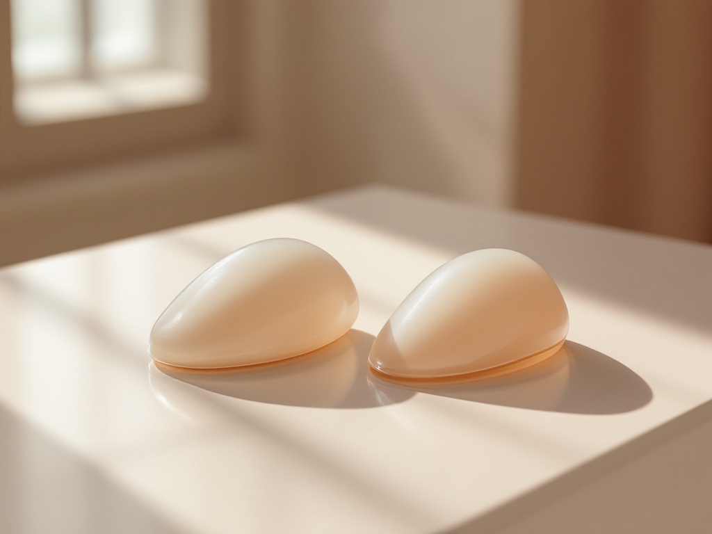
Ask three surgeons where a breast implant should sit and you may get three confident answers. That is not because anyone is wrong. It is because pocket placement is a trade-off exercise, and the right answer depends on tissue thickness, activity level, and what a patient is willing to accept over a decade or more. This explainer walks through the three main options, what each one does mechanically, and why the industry has largely converged on a hybrid approach for many patients.
The anatomy in one paragraph. The breast gland sits on top of the pectoralis major, the fan-shaped chest muscle. An implant can be placed above that muscle, directly behind the gland (subglandular), fully or mostly beneath the muscle (submuscular or subpectoral), or in a split arrangement where the upper portion of the implant sits under muscle and the lower portion sits under gland only (dual plane). A fourth variant, subfascial placement, puts the implant under the thin fibrous sheet covering the muscle but not under the muscle itself.
Subglandular: the most direct route. Placing the implant behind the gland means the muscle is untouched. Recovery tends to be faster and less painful, and there is no risk of animation deformity, the visible distortion that can occur when the pectoralis contracts over an implant. The implant also follows the natural breast footprint more closely, which can suit patients with mild sagging. The trade-off is coverage. Only skin and gland sit between the implant and the outside world. In thinner patients this raises the odds of visible rippling, a palpable implant edge, and an upper pole that looks rounded rather than sloped. Historical data also associated subglandular placement with higher capsular contracture rates, particularly with older smooth silicone devices, though modern implant surfaces and technique have narrowed that gap.
Submuscular: more padding, more compromise. Sliding the implant under the pectoralis adds a layer of living tissue over the upper portion of the device. Mechanically, this softens the transition at the top of the breast, hides ripples, and makes the implant harder to feel. Radiologists also tend to prefer it, since muscle coverage can make mammographic imaging of breast tissue somewhat easier to interpret. The costs are real, though. Dissection under the muscle means a sorer recovery, typically an extra week or two of restricted upper body activity. And because the muscle remains functional, flexing it presses on the implant. In some patients this produces visible animation deformity during workouts. Fully submuscular placement, where muscle and fascia cover the entire device, is now uncommon in cosmetic surgery because it can push the implant too high and flatten the lower breast.
Dual plane: the current default for many surgeons. Dual plane placement releases the lower attachments of the pectoralis so the muscle covers only the upper half or two thirds of the implant. The lower pole of the implant sits directly behind the gland. The logic is straightforward: keep muscle coverage where it matters most for a natural upper slope and ripple camouflage, but let the implant expand the lower breast without the muscle compressing it. This matters most in patients with mild droop or a tight lower pole, where a fully submuscular implant can ride high while the natural breast slides down in front of it, a look sometimes called a waterfall or double bubble effect. Dual plane technique exists in graded variations depending on how much muscle is released, which is why two dual plane surgeries can look quite different.
Subfascial: the middle path with a smaller evidence base. Subfascial placement aims to gain a thin extra layer of coverage without disturbing the muscle. Proponents report less animation risk than submuscular approaches with slightly better edge concealment than pure subglandular placement. The fascia is thin, however, typically under a millimeter in the upper chest, so its camouflaging power is modest. It remains a reasonable option for athletic patients with adequate soft tissue who want to avoid muscle dissection.
How the decision actually gets made. A pinch test at the upper breast is the classic screening tool. If a surgeon can pinch less than roughly two centimeters of tissue, most will steer toward some form of muscle coverage, because the implant will otherwise define the breast contour on its own. Patients who lift heavily or compete athletically may weigh animation deformity more seriously and accept a subglandular or subfascial plan with a smaller or more cohesive implant. Patients with thin tissue and a desire for larger volume are the group where submuscular coverage earns its recovery cost.
The honest bottom line. No pocket is free. Subglandular trades coverage for comfort and motion independence. Submuscular trades recovery and animation risk for concealment. Dual plane splits the difference and has become the workhorse for that reason, but it demands more surgical judgment, not less. The most useful question to ask in a consultation is not which pocket is best, but why this pocket, for this tissue, with this implant. A specific answer to that question tells you more than any brochure.
Related reading: Over, Under, or In Between: How Implant Pocket Placement Actually Works.