
Explainer · July 29, 2026 · 5 min · By Beatriz Holmgren
Over, Under, or In Between: How Implant Pocket Placement Actually Works
Subglandular, submuscular, dual plane, subfascial: the four pocket options explained by anatomy and mechanism, not marketing.
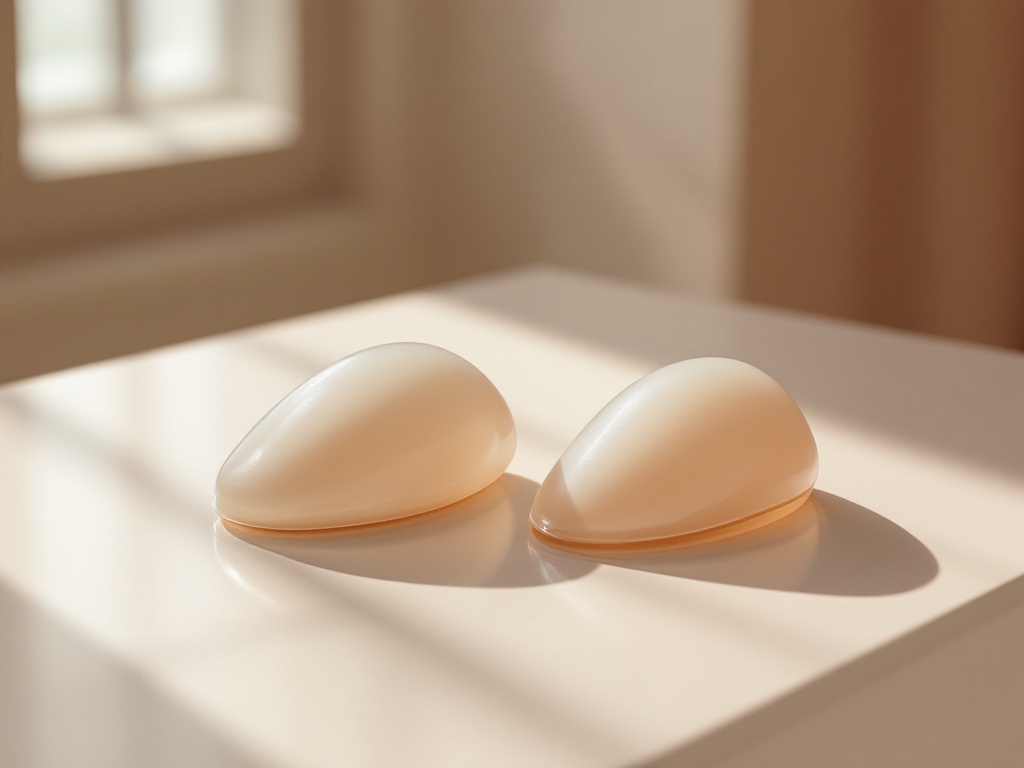
Ask three surgeons where a breast implant should sit and you may get three confident answers. That is not because anyone is wrong. It is because pocket placement is a set of trade-offs governed by anatomy, tissue thickness, and how the pectoralis major muscle behaves over time. This explainer walks through the four main options and the mechanisms behind each one, so the conversation in a consultation makes more sense.
The anatomy in one paragraph. The breast gland sits on top of the pectoralis major, a fan shaped muscle that runs from the collarbone and sternum to the upper arm. Covering the muscle is a thin connective tissue layer called the pectoral fascia. An implant can be placed above the muscle and under the gland (subglandular), under the fascia but above the muscle (subfascial), partly under the muscle (dual plane), or fully under the muscle and adjacent tissues (total submuscular, now rare in cosmetic augmentation).
Subglandular: the direct approach. Here the implant sits behind the gland and in front of the muscle. The mechanical logic is simple: the implant pushes the breast forward from directly behind it, so the result tends to look like a larger version of the existing breast. Recovery is typically faster because no muscle is divided or elevated. The trade-off is coverage. If the patient's soft tissue is thin, particularly in the upper pole, the implant edge can become visible or palpable, and rippling shows more readily with lower fill or thinner shells. Most surgeons use a pinch test at the upper breast: if the tissue pinch is under roughly two centimeters, subglandular placement raises the risk of visible implant contour. There is also a body of evidence associating subglandular placement with somewhat higher capsular contracture rates, possibly because the pocket sits closer to ductal tissue and its bacterial flora, which is one proposed driver of capsule inflammation.
Submuscular and the dual plane compromise. Placing the implant under the pectoralis adds a layer of living, vascular tissue over the upper half of the device. That extra coverage softens the transition at the upper pole, reduces visible rippling, and appears to lower capsular contracture rates in most published series. Mammography visualization is also generally considered easier with the implant behind the muscle. The cost is muscular. Dividing the lower attachments of the pectoralis means more early postoperative discomfort, and the muscle keeps working for life. When the patient contracts the chest, the implant can visibly shift or distort, a phenomenon called animation deformity. It ranges from trivial to genuinely bothersome, and it matters more to people who lift weights or perform on stage.
The dual plane technique, described in the early 2000s and now the most common approach in many practices, splits the difference. The upper portion of the implant sits under muscle for coverage, while the lower portion sits under gland, allowing the implant to fill the lower pole and letting the gland redrape over it. Surgeons adjust how high the muscle is released to match the patient's tissue: more release for droopier or more constricted breasts, less for tight youthful tissue. Mechanistically, dual plane exists because the upper breast needs coverage and the lower breast needs expansion, and one pocket rarely serves both goals equally.
Subfascial: the newer middle path. The pectoral fascia is thin, often about half a millimeter, but it is a distinct structural layer. Lifting it and placing the implant beneath it keeps the muscle intact, so there is no animation deformity, while the fascia adds a modest degree of support and edge smoothing compared with a purely subglandular pocket. Advocates report faster recovery than submuscular placement with better upper pole blending than subglandular. Skeptics note that a half millimeter layer cannot substitute for muscle in a very thin patient, and long term comparative data remain limited relative to the older techniques. The honest summary is that subfascial placement works best in patients with moderate tissue thickness who want to avoid muscle involvement.
What actually drives the decision. Four variables do most of the work: soft tissue thickness (the pinch test), the patient's activity profile (heavy chest training favors avoiding the muscle or accepting animation trade-offs), the degree of sag (more ptosis often pushes toward dual plane or a lift), and implant characteristics (larger or lower fill devices need more coverage). No plane is universally superior. A plane that is ideal for a thin patient with minimal breast tissue may be unnecessary for a patient with generous natural coverage.
Questions worth asking in consultation. Which plane do you recommend for my tissue thickness, and what did my pinch test show? If dual plane, how much muscle release do you plan and why? What does animation deformity look like in your patients, and how often do they mention it? How would this plane affect a future revision? A surgeon who answers in terms of your anatomy, rather than a single preferred technique for everyone, is reasoning the way this decision deserves.
Related reading: Over, Under, or In Between: How Implant Pocket Placement Actually Works.